























Week of 04-Sep-2026
Several growing and finishing pigs vomit after eating and gradually lose body condition. After a few days they are euthanized. The only gross post-mortem lesion is a megaesophagus. What is cause of this lesion in these pigs?
Megaesophagus is characterized by dilation and general increase in size of the esophagus, absence of peristalsis, presence of tertiary contractions and partial or total inability of the gastroesophageal sphincter to relax. These changes could lead to dysphagia that, consequently could compromise the pig’s nutritional state. In addition to regurgitation and weight loss, coughing, mucoid and purulent nasal discharge, as well as dyspnea with pneumonia due to concomitant aspiration may be observed. Regurgitation at variable frequencies and weight loss are among the clinical signs observed in the animal. The diagnosis made for these animals was based on observation of clinical signs and post-mortem examinations. Post-mortem examinations of all pigs revealed ulceration of the pars esophagea of the stomach. When the ulcer heals, it leads to scarring or, in severe cases, to strictures, as seen in these cases. The stenosis of the esophageal opening appears as a rigid ring, and the opening can become so small that pigs often vomit after eating, as seen in this case. Different factors contribute to the ulceration of the stomach. These could be infectious, nutritional, behavioral and/or management-related factors.
Treatment in cases like this are usually supportive or surgical procedures. Supportive treatment is characterized by changes to animal management practices, in addition to the utilization of drugs such as antibiotics, prokinetics, and antacids. Surgical methods can be performed using several techniques, such as the esophagus-diaphragmatic cardioplasty, or Heller myotomy, associated with fundoplication (none of these are practical in a farm setting). Some animals with congenital idiopathic megaesophagus are able to recover on their own while the acquired condition is irreversible. In regards to secondary megaesophagus, once the cause of the problem is resolved, the animal has a chance of recovering. Therefore, prognoses for acquired and congenital megaesophagus range from guarded to favorable, while for the acquired cases it is poor.
Photo of the week
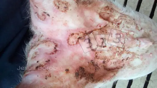
Week of 16-Oct-2026
This weaner pig has non-pruritic lesions on the skin. What is the cause of this lesion?
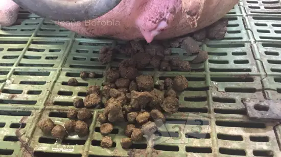
Week of 09-Oct-2026
Small, pellet-shaped portions of manure are seen behind a sow after farrowing. What is the cause of this condition?
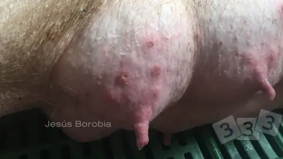
Week of 02-Oct-2026
Some damage to the teats is found when inspecting the udder of this sow. What is the cause?
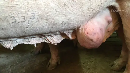
Week of 25-Sep-2026
A sow is seen with a firm mass in the mammary glands. What is this condition?
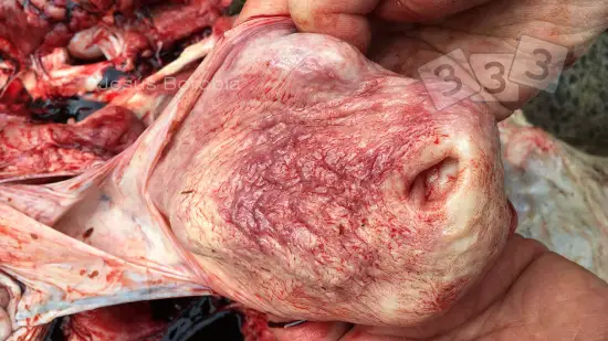
Week of 18-Sep-2026
What is the name of this lesion in the bladder?
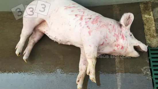
Week of 11-Sep-2026
A finishing pig is found in the pen lying on one side and gasping for air, with skin discoloration and muscle tremors. What is the most likely cause of this problem?
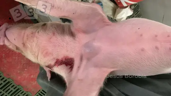
Week of 28-Aug-2026
When a piglet was handed for blood testing, a lump was detected on its sternal area. What is this condition?
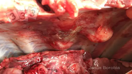
Week of 21-Aug-2026
In the post-mortem of a gilt, some nodules are found in the chest cavity. What are they?
Week of 14-Aug-2026
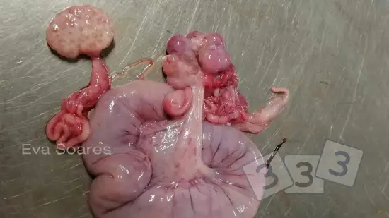
Ovaries obtained from a female fattening pig at the slaughterhouse. The ovary on the left side of the image is normal but the ovary on the right side and uterus have some abnormalities. What could be the cause?
Week of 07-Aug-2026
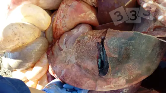
Which of the following lesions do you see pictured?
Week of 31-Jul-2026
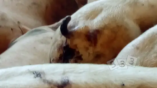
We notice dark diarrhea in finishing hogs. What could cause this?
Week of 24-Jul-2026
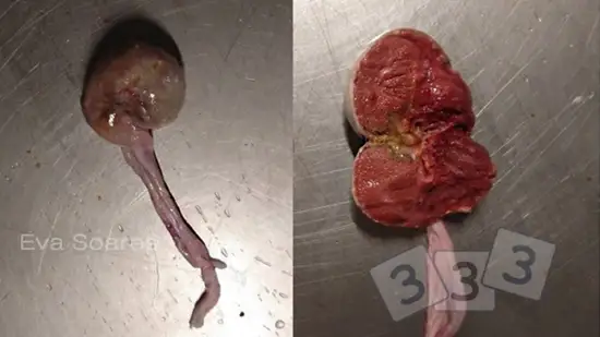
During meat inspection at the slaughterhouse, two globular masses were found in the ovarian insertion. After cutting the mass, its internal structure was similar to that of normal testicles. What is the name of this developmental abnormality?
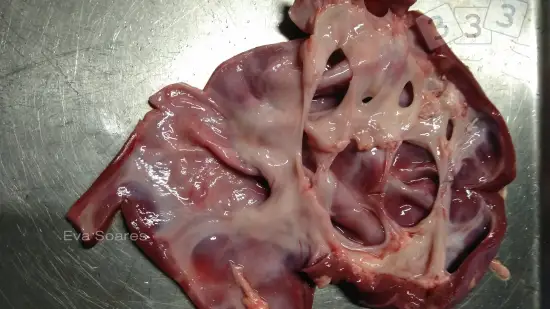
Week of 17-Jul-2026
Lesion found at slaughter in the kidney of a fattening pig. What is this pathology compatible with?
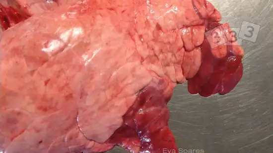
Week of 10-Jul-2026
Red areas of cranioventral consolidation (hepatization) were detected in this lung at the slaughterhouse, suggesting enzootic pneumonia. What is the main agent associated with this condition?
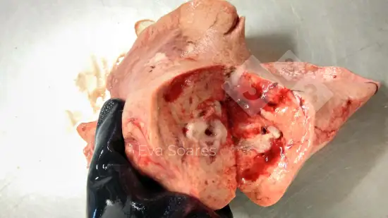
Week of 03-Jul-2026
Lung at the slaughterhouse with foam mixed with fluid. What is the most likely cause?
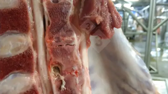
Week of 26-Jun-2026
This suppuration was observed in the spinal column during the post-mortem slaughterhouse inspection of a pig carcass. What is it?
Week of 19-Jun-2026
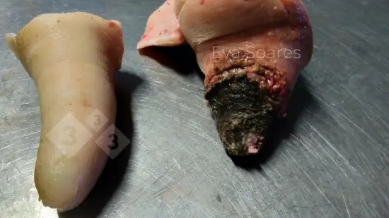
Pictured on the left is an uninjured tail from a finishing hog, and on the right side a tail that suffered tail biting. What is the most common cause of this problem?
Week of 12-Jun-2026
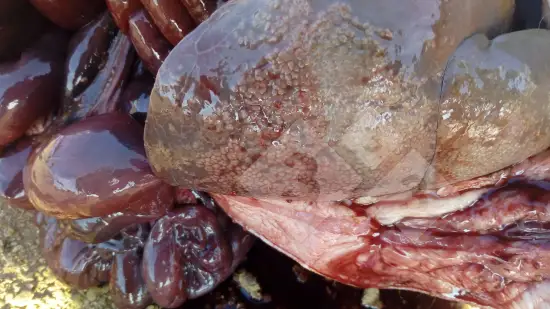
Does the presence of this type of lesion always imply a disease problem?
Week of 05-Jun-2026
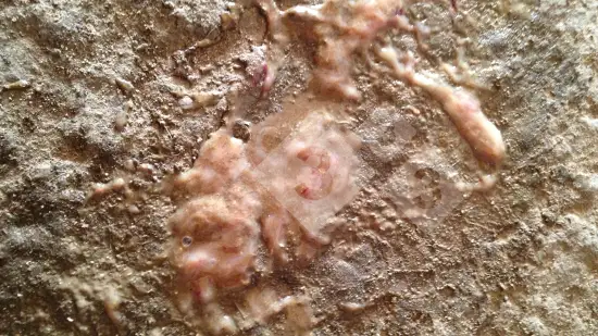
In a pen of growing pigs that had stopped eating, this kind of feces is seen on the floor. What could be the cause?
Week of 22-May-2026
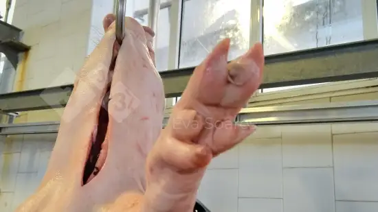
This pig has an anomaly on its foot. Do you know what it is?
Week of 15-May-2026
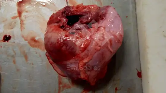
In the slaughterhouse inflammation of the parietal and visceral face of the pericardium was found in a finishing hog. What is the name of this lesion?
Week of 08-May-2026
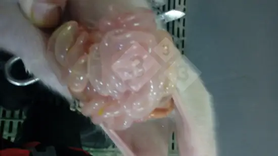
Litters that are 3 days old show vomiting and diarrhea that lasts two or three days. What could be the cause?
Week of 24-Apr-2026
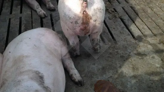
A pen of fattening pigs stopped eating and have lost body condition. This pig's perianal area is dirty. What could be the cause?
Week of 17-Apr-2026
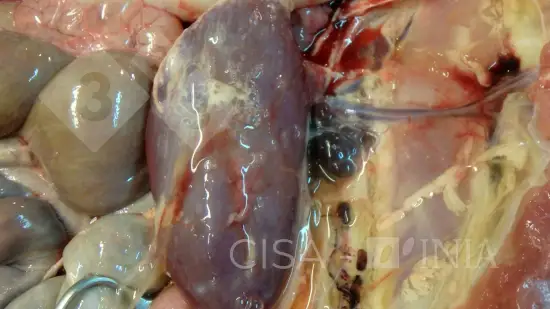
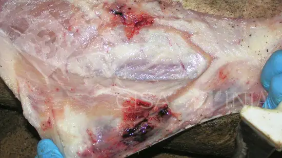
What could be the cause of this lesion?
Week of 10-Apr-2026
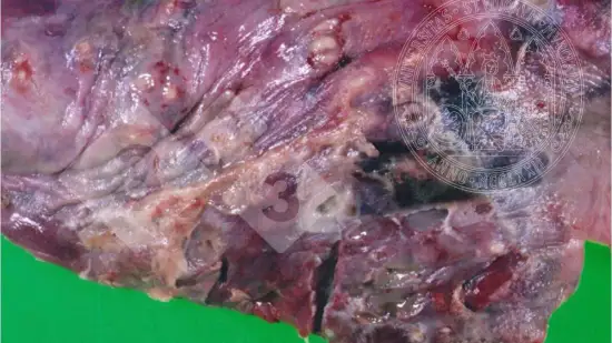
What disease could cause those lesions?
Week of 03-Apr-2026
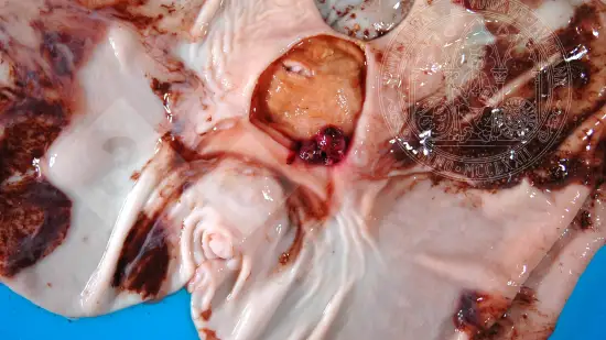
What is the most probable etiology of this lesion?
Week of 27-Mar-2026
The origin of this lesion is?