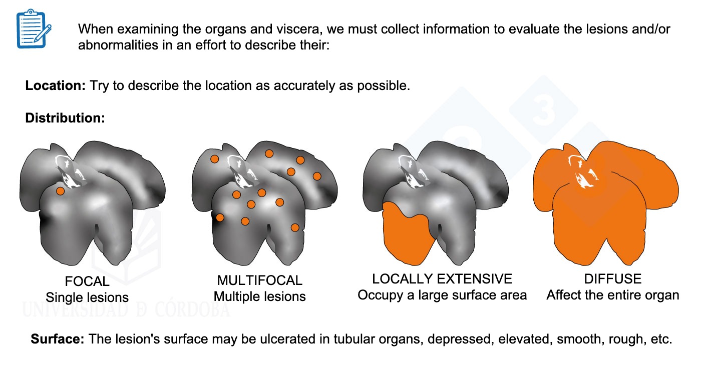
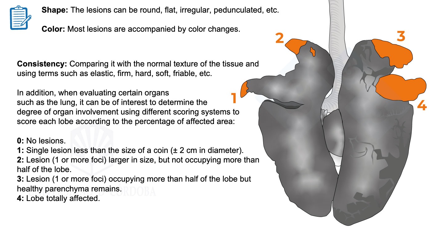
When examining the organs and viscera we must gather information to evaluate the lesions and/or abnormalities. We will learn to describe their location, distribution, surface, shape, color, and consistency.
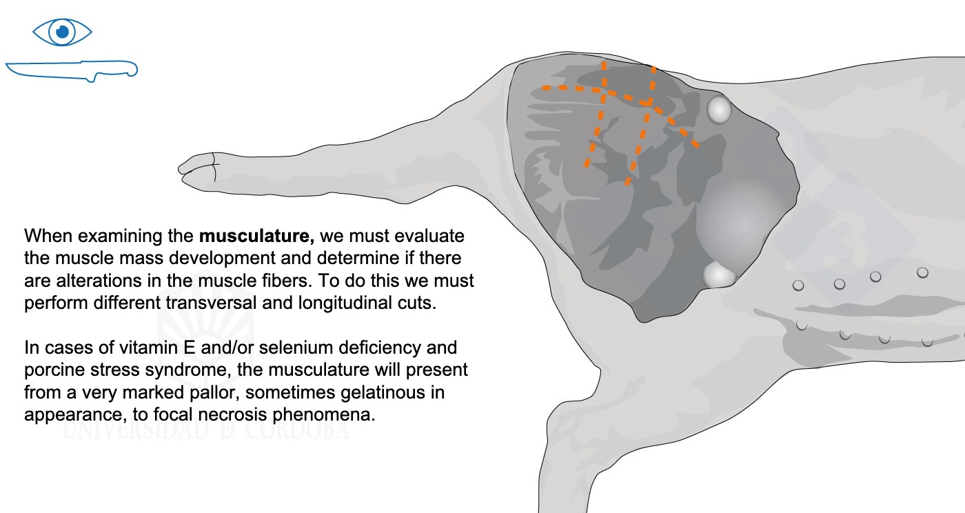
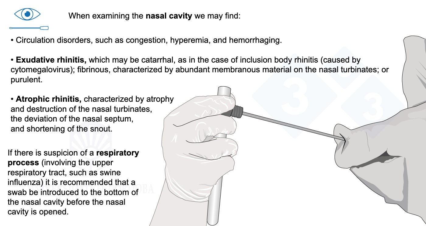
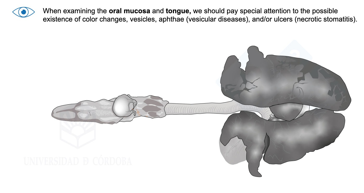
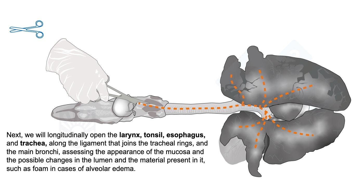
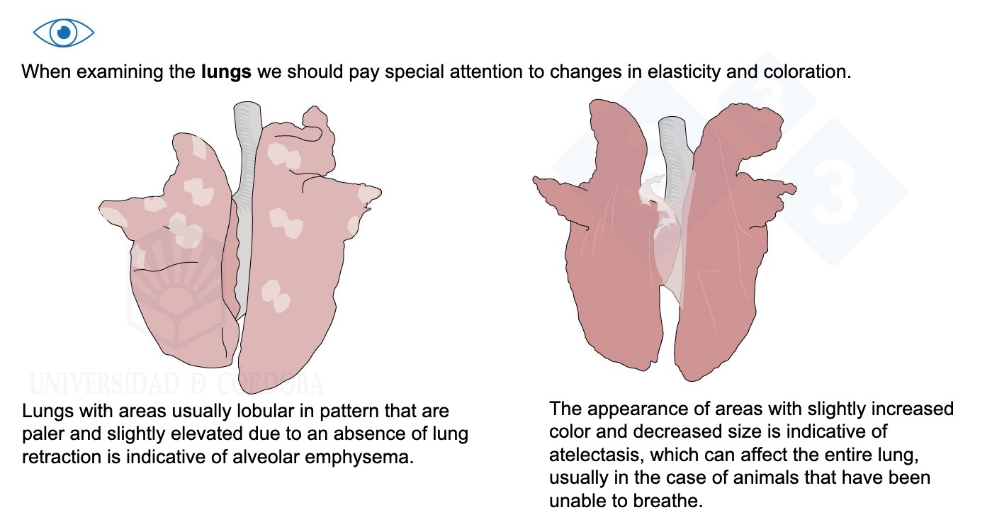
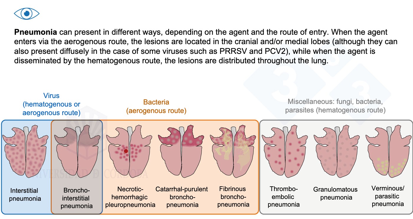
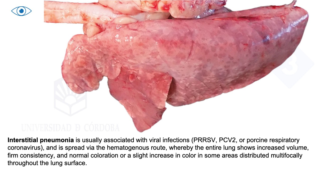
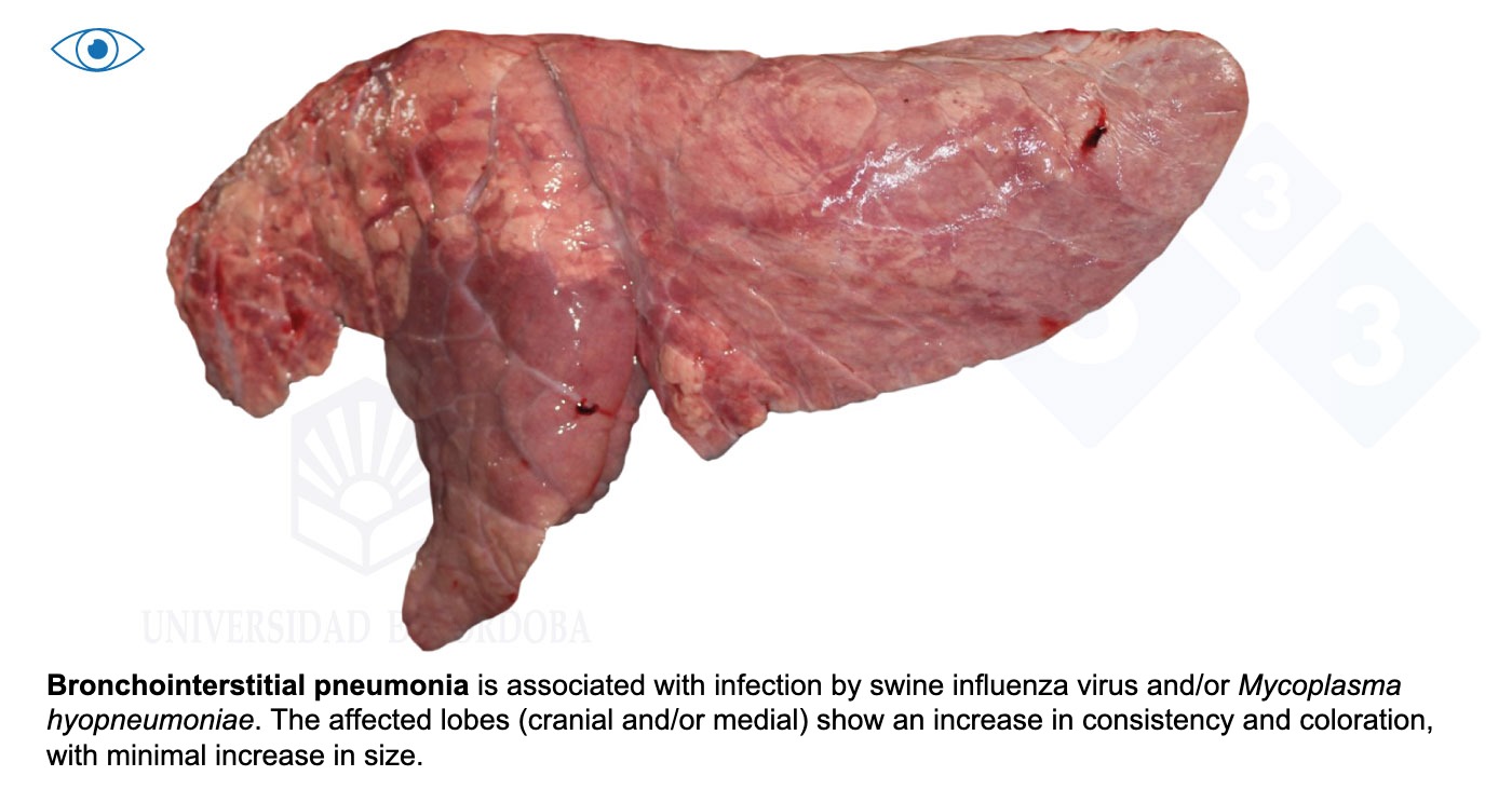
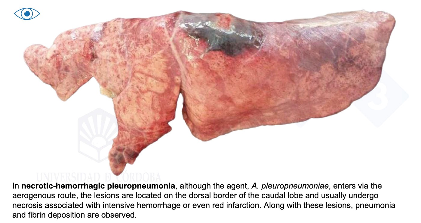
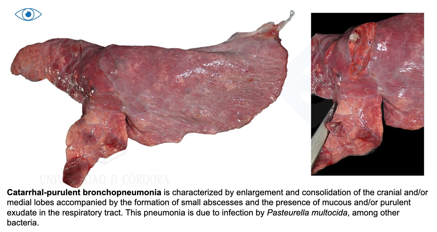
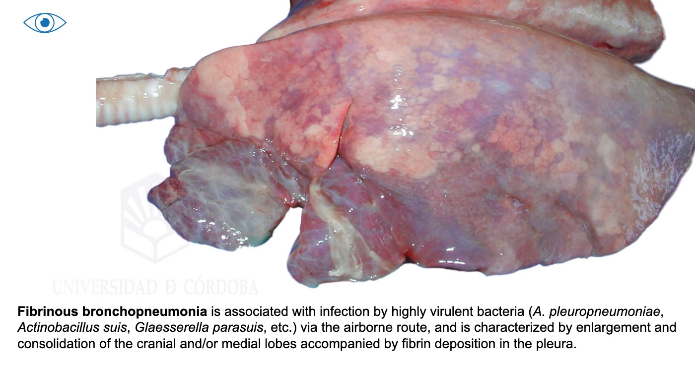
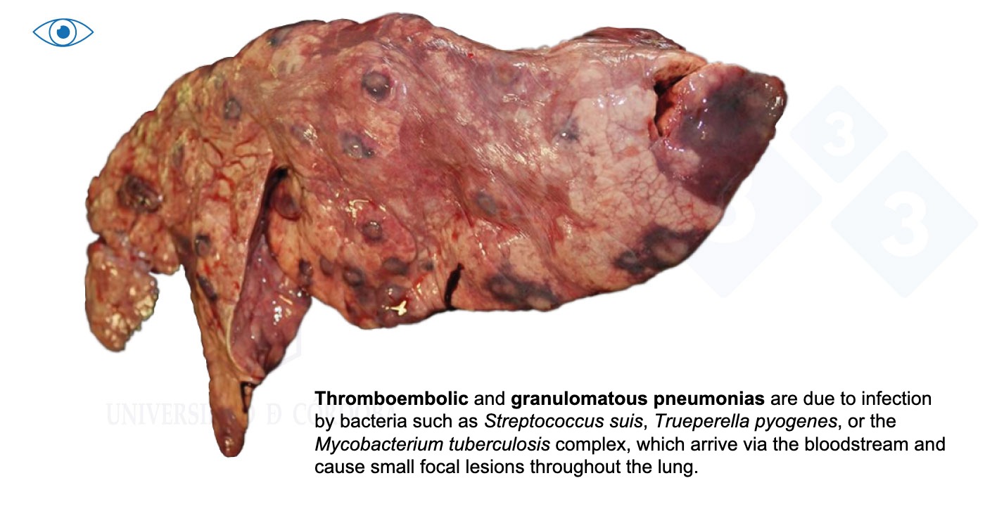
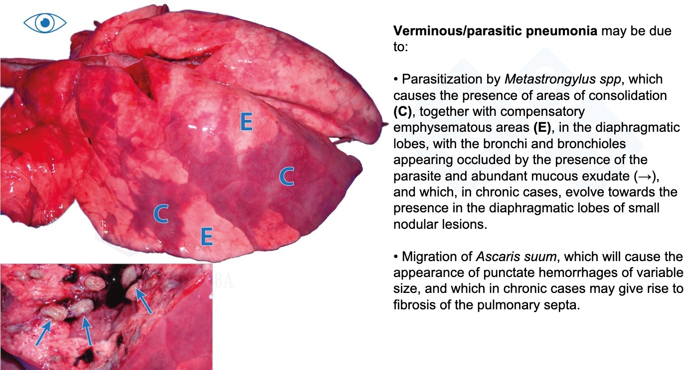
After examining the musculature and joints, we will examine the nasal cavity, oral mucosa, tongue, larynx, tonsils, esophagus, trachea, and main bronchi. In the examination of the lungs, we will address how to differentiate between different types of pneumonias with real photos.

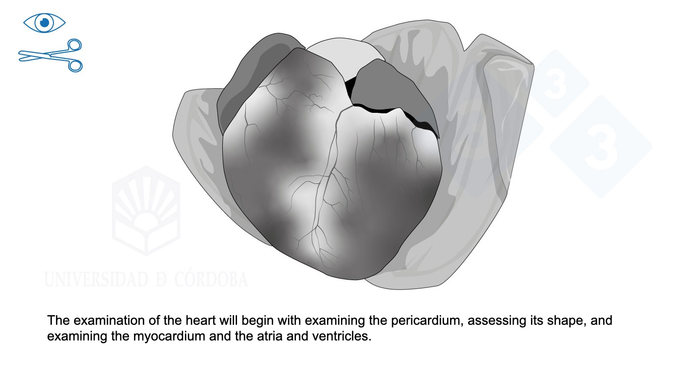
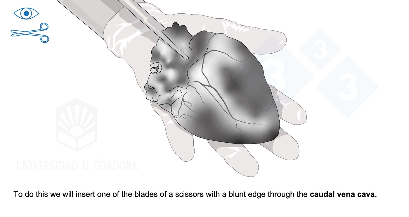
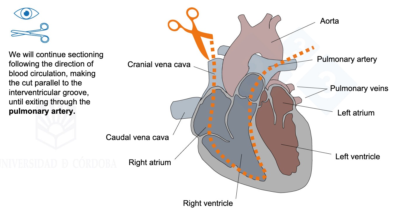
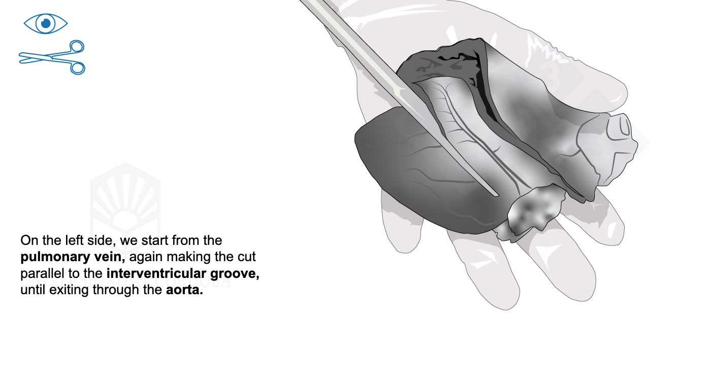
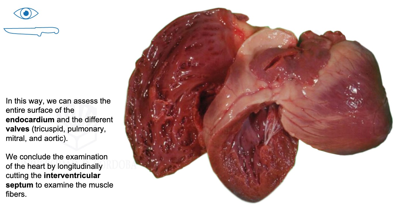
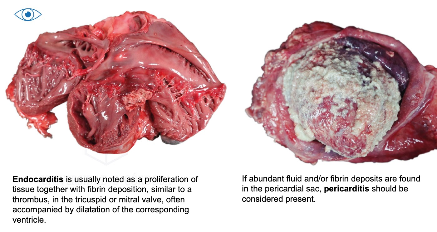
Then we will show the technique of opening the heart to examine the pericardium, myocardium, atria, and ventricles, and we will see photo examples of endocarditis and pericarditis.