Digestive pathologies at the beginning of the growing phase: same bacteria and two different problems
02-Sep-2021 (4 years 10 months 14 days ago)
1. Introduction
The most critical phase of the finishing period is the first month. How the first days start off will determine the finishing performance. It is in this period when digestive issues normally occur, pathologies that are usually very aggressive, causing a large number of losses in a short period of time, plus subclinical disease in the rest of the animals that will result in reduced production indexes.
We are going to focus on two types of pathologies caused by two different pathotypes of Escherichia coli.
- Edema disease
- Enterotoxigenic colibacillosis
2. Pathogenesis
In both cases, the bacteria need to bind via the F18 fimbriae to avoid being swept away by intestinal motility. Receptors for these fimbriae develop in piglets starting at weaning, a fact that determines that during lactation we do not find these digestive issues.
2.1. Edema disease:
The Shiga toxin (Stx2e) is responsible for the clinical signs and lesions. It crosses the intestine and binds to erythrocytes, causing significant vascular damage that increases vascular permeability, leading to edemas in different parts of the body (brain, eyelids, face, larynx, mesocolon), leading to rapid death in the animals.
2.2. Enterotoxigenic colibacillosis:
The release of LT (heat-labile) and ST (heat-stable) toxins is responsible for the clinical signs as they alter intestinal homeostasis and produce hypersecretion of fluids and electrolytes into the intestinal lumen. LT produces the opening of the anion channels and relaxes the junction between enterocytes causing a secretion of chloride and bicarbonate ions into the intestinal lumen, converting the intestine into a hypertonic medium. This causes the outflow of water from within the cells in an attempt to balance the ion concentration, leading to the onset of diarrhea. The effect of LT once initiated is irreversible.
 ST inhibits the absorption of ions. The association of both toxins generates dehydration and metabolic acidosis in piglets.
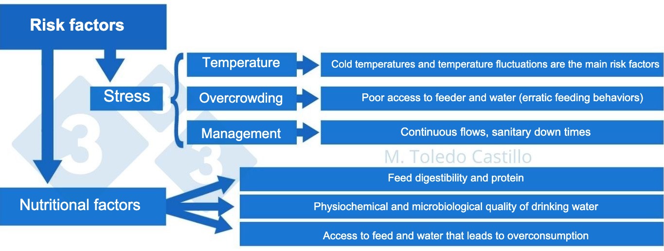
3. Predisposing factors
- Stress. The release of cortisone reduces the immune response.
- Concomitant viral infections.
- Changes in the normal microbiota of the intestine caused by changes in feed or poor water quality can affect the maintenance of the intestinal barrier. The epithelial cells of the intestine form a semi-permeable barrier that, under normal conditions, prevents the passage of antigens and toxins. During inflammatory phenomena in the intestine (e.g. e.coli related diseases), the integrity of the intestinal permeability is lost, allowing the entry of pathogens.
- Antibiotic treatments that reduce competitive inhibition with other bacterial agents and decrease the diversity of the intestinal microbiota providing an opportunity for pathogenic microorganisms to grow in the intestine.
- Changes in intestinal motility. The decrease in intestinal peristalsis allows the proliferation of bacteria in the intestine since the efficacy of one of the mechanisms that favor their elimination is reduced.
- Low temperatures decrease intestinal peristalsis and, therefore, decrease the digestive cleansing and dragging mechanism. Large variations between daytime and nighttime temperatures favor the appearance of these pathologies.
- Protein concentration in the feed. Protein that reaches the intestine undigested serves as a substrate for bacterial growth.
- Quality of drinking water. The incidence is higher in water with higher conductivity and alkaline pH.
- Withdrawal of zinc oxide produces a change in the pattern of occurrence of edema disease. Zinc oxide does not prevent the proliferation of E. coli, but it does inhibit the production of the toxin in the bacterial cytoplasm. Since it is an endotoxin, the bacterial death due to the use of antibiotics causes the endotoxin to be released and influences the stability of the enterocyte membranes to fluid loss. Since the withdrawal, certain pathologies that were kept hidden have increased in prevalence.
 4. Clinical signs
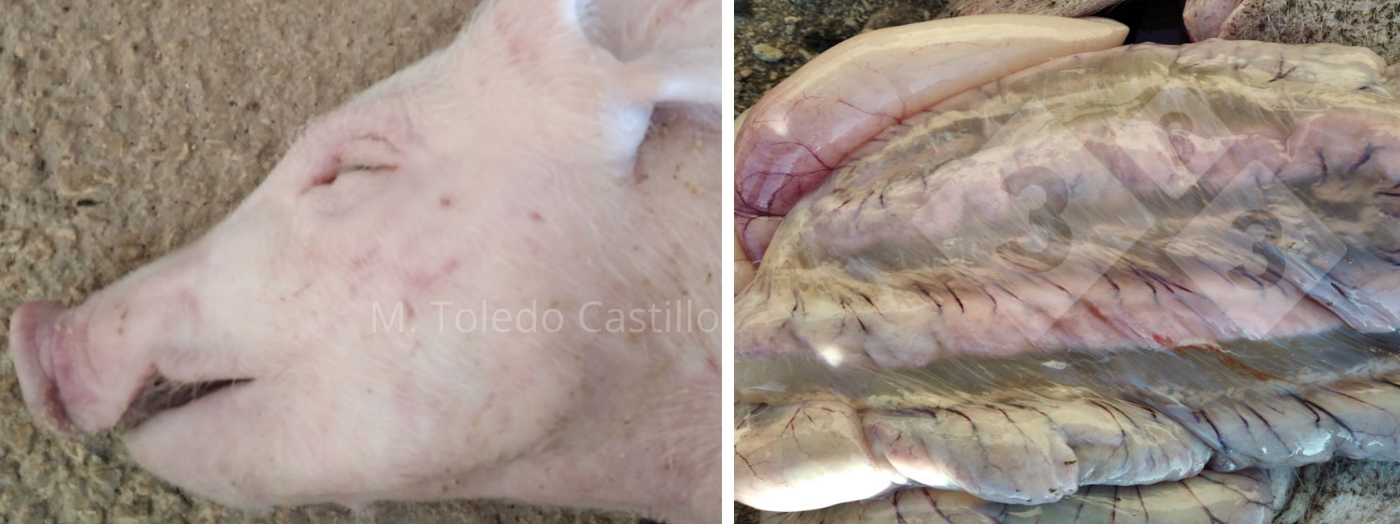
4.1. Edema disease
Edema at the brain level causes a lack of coordination, staggering gait, or the inability to walk in the absence of fever. We can often observe edema on the face, especially on the eyelids. Animals present respiratory distress as a consequence of edema and vascular damage of the airways (video). Laryngeal edema produces a specific grunting sound. The strongest piglets in the barn are usually affected. Death ensues extremely rapidly in a large number of animals. Gelatinous edema is observed in the colon and bloody edema in the tissues. We can find petechiae in the intestine and an excess of serous fluid in the abdominal cavity.
4.2. Enterotoxigenic colibacillosis:
The animals have sunken eyes due to severe dehydration. Piglets have sunken flanks. Sometimes there is severe septicemia and areas of cyanosis. Death is due to dehydration and metabolic acidosis. Necropsy shows a very congestive intestine with a large amount of liquid inside. This intestinal content is often hemorrhagic.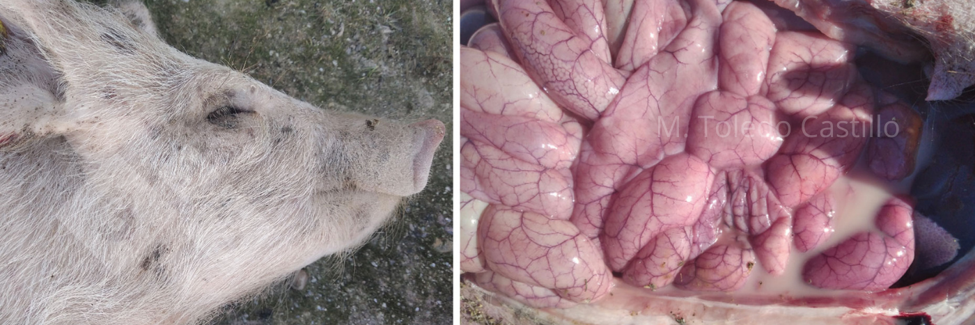
5. Diagnosis
Clinical signs and necropsy lesions allow us to establish a correct clinical diagnosis, but a laboratory diagnosis is necessary for confirmation and to identify the virulence factors of the E. coli involved:
- Identification of fimbriae, the adhesion factor to the intestinal epithelium, without which they cannot exert their pathogenic effect.
- Identification of the toxins, which produce the damage.
Identifying virulence factors will define the pathotype of the E. coli we are facing and then MICs (minimum inhibitory concentrations) are requested to know which antibiotic is the one of choice.
6. Disease control - General measures
- Cleaning and disinfection of the floor, walls, and other equipment, including feeders and drinkers. Allow the barn to dry. Verify that cleaning and disinfection are carried out correctly and efficiently.
- Cleaning of the water pipes i including the section going to each of the drinker nipples.
- Monitoring of drinking water: checking microbiological and physicochemical quality.
- Perform a good sanitary down time, with cleaning of the slurry pits, if possible.
- Empty the silos to avoid any feed left over from the previous batch. Clean and disinfect silos to eliminate possible mycotoxins.
- Reduce the protein level of the initial diet. Feed protein can be reduced until pigs reach 40 kg without major production losses, as clinical and subclinical presentation is reduced.
- The temperature of the barn when the piglets arrive should be around 22ºC. If necessary, we will use systems to heat the barn and try to fill the barn in the morning. If piglets arrive at the barn in the afternoon, a temperature above 18ºC will not be reached until the following day. Install green house type of covers (see photo) that reducesthe volume of air to be heated. Complement with a heat source (heating systems or portable forced air heaters with a thermostat).

- Avoid the use of bactericidal antibiotics for the control of other pathologies since they cause dysbiosis and predispose pigs to these digestive problems.
7. Disease control - Specific measures
- Vaccination with the edema disease toxoid is extremely effective in controlling the disease in the finishing period. The problem is that on many occasions we observe that if TL and ST toxins along with shiglatoxins (several pathotypes at the same time) appear in the analysis, we can control edema disease, but we do not know the clinical impact that the rest of the toxins can have in the presentation of enterotoxigenic colibacillosis. Vaccination can be used as a treatment since several batches of animals can be vaccinated and the effectiveness of the vaccination can be tested. Immunity is achieved 21 days after administration. The time of administration will vary depending on when the problem appears.
- Oral vaccination for the control of E. coli (F4 and F18) can be a tool for reducing enterotoxigenic processes in the nursery and at the beginning of growing/finishing. In our case, we lack the data to be able to evaluate it to its proper extent.
- Restricted feeding. This is a common practice together with the use of oral antibiotics. There may be problems if feed access is not done in a gradual way once feed restriction is over, since overconsumption can lead to resurgence.
- The use of organic acids is suitable to prevent the proliferation of bacteria in the intestine, not only because of the decrease in pH they produce but also because of their antimicrobial power.
Regarding edema disease, we can conclude that, in our case, the treatment that gave us the best results in the presence of outbreaks, has been feed restriction as a management measure, together with the administration of zinc oxide in the drinking water as there are currently several authorized products in the EU that can be used at the beginning of the growing phase, Zinc oxide is an effective treatment, but we must remember that its use will be banned in the European Union and the UK on 26th of June 2022, and therefore there will be a higher incidence of these pathologies on our farms. From this moment, the control of the disease should be carried out avoiding its emergence through the administration of toxoid vaccines.